Published Date: Sep 05 2023
Cells are important materials in biological experiments and the foundation of Biological cytology experiments. Lots of research related to drugs, gene functions and disease mechanisms, etc. needs to be explained at the cellular level. The operation process of cell culture and detachment is greatly influenced by personnel operation and environmental conditions which, any one of the factors, may result in a failure of the process.
Hope the following notices about cell culture and digestion summarized by biological pharma company GeneBicon will help the researcher who is operating cell culture and cell detachment.
1. Cell line identity needs to be clarified.
The reason why a certain cell line is selected for the experiment is that some characteristics of the selected cell line are in line with the setting of the research direction, for example, tissue characteristics, disease characteristics, functional characteristics, gene expression characteristics, etc. Once the wrong cell line was selected would lead to large errors and uncertainties.
Resent years, several authoritative organizations have found serious cross-contamination of cell lines during culture and circulation. According to incomplete statistics, the pollution caused by the HeLa cell line alone has caused problems in the accuracy of the results of more than 30,000 papers (doi:10.1371/journal.pone.0186281). And this situation is not limited to HeLa cells. Researchers from multiple authoritative institutions have found that more than 451 cell lines have been completely absorbed by other cells, and the contamination accounted for 46% of the tested cells. It can be seen that the phenomenon of cell cross-contamination is very common. Therefore, it is very important to verify the identity of the cell line before doing the experiment. If the cell line is purchased from an institution, it is best to ask the supplier to provide a qualified STR identification report.
2. Cell culture and Cell Detachment
Cell Culture
Normally, there are tow kinds of cell culture, one is suspension cell culture and the other is adherent cell culture. Either way, the following steps are generally followed for cell culture:
Cell preparation↓Cell passage↓Cell medium configuration↓Cell inoculation↓Culture condition control↓Cell observation and culture↓Cell experiment↓Cell cryopreservation
Of cause, there is a basic framework of the cell culture process above and the practical operation may vary from the type of cell, experiment request and lab’s standard operation procedures.
Cell Detachment
Cell detachment is a process of operating cell culture by R&D. Generally when the cells grow to 80%-90% density, they need to be subcultured and detached. Prior to subculturing adherent cells, it is necessary to use a digestion reagent to dissociate the cells from the culture container surface. The cells must then be separated into a single suspension and transferred to a new container filled with fresh medium. There are three common cell detachment methods: enzymatic digestion, ion chelation, and mechanical digestion.
Among the three common cell detachment methods, enzymatic digestion is the most widely used method, mainly using trypsin, the general concentration is 0.25-0.5%, and the digestion time varies according to factors such as cell type and action temperature. In general, at 37°C, 1-5 minutes is enough for 0.25% trypsin digesting a monolayer of adherent cells. It should be noted that Ca2+, Mg2+, serum, and protein can reduce the activity of trypsin, so trypsin without Ca2+ and Mg2+, and EDTA-containing trypsin has higher activity; At last, trypsin digestion can be terminated with serum-containing medium.
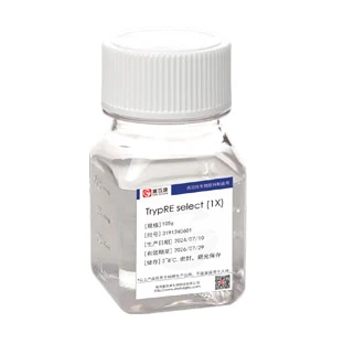
There are roughly 2 kinds of trypsin, one is traditional animal-origin trypsin mainly extracted from bovine or pig pancreatic tissue whose disadvantage is the risk of viral contamination, another is animal-free trypsin which is produced by gene recombination technology. Recombinant enzymes have many potential usages including drug production, application of biotechnology or as a research tool, for example, protein drug production, cell segregation, vaccine production and tissue sample preparation for cell analysis while avoiding the risk of viral contaminant brought by bovine and pig pancreatic trypsin. 2020 edition of China Pharmacopeia includes for the first time the quality criteria for non-animal-derived recombinant trypsin. After all, recombinant trypsin is an indispensable tool enzyme in the field of biological medicine and scientific research.
3. Steps for the use of recombinant trypsin in practical applications:
A. Preparing buffer: Preparation of 10L HBSS balanced salt solution with sterilized water.
The HBSS balanced salt solution formula: 400mg/L KCl,60mg/L KH2PO4,350mg/L NaHCO3,8000mg/L NaCl,121mg/L Na2HPO4·12H2O,1000mg/L glucose.
B. Weighing 1g recombinant trypsin (cell culture grade), dissolved and clarified with HBSS balanced salt solution to get a trypsin enzyme activity concentration of 500USP units/ml-1500 USP units/ml, or to make the trypsin concentration of 0.1mg/ml-0.3mg/ml. (The concentration of 500USP units/ml is suitable for most cell detachment, and it can be increased to 1500 USP units/ml for the primary cell and tissue detachment.
4. Notifications:
1. Gene-Biocon Recombinant Trypsin Detach Solution contains EDTA which determines the adaptability to different cells. In addition, this solution should be used in a sterile environment as it does not contain bacteriostatic agents and may become contaminated by microorganisms.
2. It is suggested that the recombinant trypsin detach solution be divided in 100ml/vial and stored at -15℃ ~ -20℃ which can be stored stably for 12 months. It is not suitable for long-term storage at 4°C. Avoid repeated freezing and thawing to reduce the activity of trypsin and reduce the probability of contamination.
5. Q&A
What is EDTA? Why do we add EDTA in recombinant trypsin?
EDTA (Ethylenediaminetetraacetic acid, also called edetic acid after its own abbreviation), is an amino polycarboxylic acid with the formula [CH2N(CH2CO2H)2]2. This white, water-soluble solid is widely used to bind to iron (Fe2+/Fe3+), magnesium (Mg2+) and calcium ions (Ca2+), forming water-soluble complexes even at neutral pH. This ions are important factors in maintaining organizational integrity. But EDTA alone cannot disperse the cells completely, so it is often mixed with trypsin in different proportions, and the effect is better. The usual ratio is 1:1 or 2 parts EDTA:1 part trypsin. The working solution concentration of EDTA is 0.02%, and it is prepared with BSS without Ca2+ and Mg2+. If EDTA is used, it is necessary to remove the digestion solution with buffer before adding culture solution before terminating the digestion.
How to control the digestion time of trypsin?
The detachment times vary from the sensibility to the detach solution of cells. The trypsin detachment time is related to the trypsin concentration, whether it contains EDTA, the storage time and temperature of trypsin, whether it’s repeatedly frozen and thawed, the volume of trypsin added for detachment, the detachment temperature and the density of cells. For the detachment of newly-purchased cells, it is recommended to use a low concentration of trypsin to carefully explore the detachment time and to observe whether the cell becomes round every 1 minute and record the optimal detachment time. The next operation can refer to the previous record to control the time.
The culture medium was stored at -20℃ by mistake, can I continue to use it after dissolving it?
At -20℃, salts in the culture medium tend to precipitate and may not redissolve when melted again which results in the variety of nutrient composition and osmotic pressure of the medium. If cells are cultured in this medium, they rupture easily. It is recommended to discard this culture medium and change to a new one.
What are the main characteristics of cellular overdigestion?
Quite parts of cell death and damage. The cell mass flows on the medium and the white membranoid substance is visible just after passage. If the cell cluster is full of dead cells, there will not be seen adherent cell. If some active cells exist, the adherent cell density will be uneven, which will quickly form a central necrotic cell area.
What happens when the cells cannot be detached?
(1) Some cells adhere more firmly to the wall, so there will always be some cells left after each detachment and pipetting, or the cells have grown well but the culture flask has not been changed for a long time. In order to protect most of the viable cells, cell digestion should be stopped in moderation.
(2) Different types of cells require different detachment times. Because the cell detachment of adherent cells usually takes about 2 minutes, it is difficult to detach individual cells. If it is still ineffective after multiple detachment and repeated pipetting, just give up.


